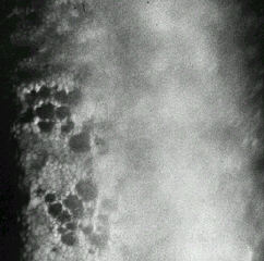
This is the specular photograph of a patient awaiting cataract surgery. The corneal thickness measures 0.65 mm with pachymeter and there is no clinical evidence of corneal oedema. What is responsible for the dark areas and what is the diagnosis? Answer The dark areas are caused by corneal guttata. The photograph shows decreased concentration of corneal endothelium. This is a case of early Fuchs' endothelial dystrophy. More questions
This is the specular photograph of a patient awaiting cataract surgery. The corneal thickness measures 0.65 mm with pachymeter and there is no clinical evidence of corneal oedema. What is responsible for the dark areas and what is the diagnosis? Answer The dark areas are caused by corneal guttata. The photograph shows decreased concentration of corneal endothelium. This is a case of early Fuchs' endothelial dystrophy.
This is the specular photograph of a patient awaiting cataract surgery. The corneal thickness measures 0.65 mm with pachymeter and there is no clinical evidence of corneal oedema.
What is responsible for the dark areas and what is the diagnosis?